Structure of IDP91202
1.60 Angstrom Resolution Crystal Structure of a 3-Dehydroquinate Dehydratase-like Protein from Bifidobacterium longum
Annotation
- Description
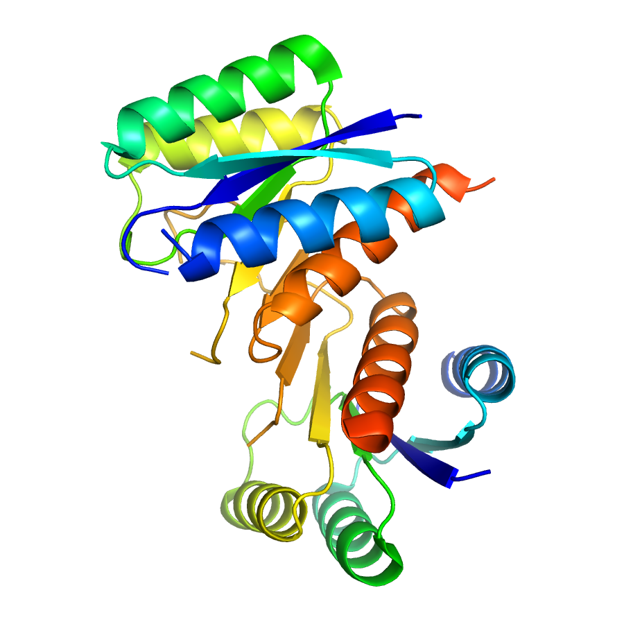
- The shikimate pathway links metabolism of carbohydrates to biosynthesis of aromatic compounds. In a sequence of seven steps, phosphoenolpyruvate and erythrose 4-phosphate are converted to chorismate, a precursor of the aromatic amino acids and many secondary aromatic metabolites. The shikimate pathway is essential for most bacteria and plants but absent in humans, making it an attractive target for the development of novel antibiotics. The third step in the pathway consists of the dehydration of dehydroquinate to dehydroshikimate. This reaction can be catalyzed by two enzyme families which utilize distinct mechanisms. While this protein is annotated as a type II dehydroquinate dehydratase and indeed has high sequence homology to active dehydratases, the structure reveals notable differences relative to all other characterized enzymes. Whereas other type II dehydratases assemble as dodecamers, comprised of a tetramer of trimers, this protein is assembles as a dimer, preserving the trimer-trimer interactions of the dodecamer. In other dehydratases a neighboring molecule in the dodecamer extends into the active site, making substantial contacts with the substrate and other active site residues. With the affected oligomeric assembly observed in this structure, comparable protein-protein interactions are noticeably lacking and much of the active site is disordered. At present, the source and functional implication of the unique these unique structural features await characterization.
- Functional assignment
- Unknown
Ligands
| Ligand code | Name | Ligand type |
|---|
Structure information
Unit cell parameters
- Space Group
- P 21 21 21
- Unit Cell
-
a=44.00Å, b=72.68Å, c=81.71Å
α=90.00, β=90.00, γ=90.00 - Solvent content
- Matthews coefficient
- Resolution range
- 29.94-1.60Å (1.64-1.60Å)
- Rall(%)
- 17.8
- Rwork(%)
- 17.6 (22.7)
- Rfree(%)
- 20.3 (26.6)
- Num. observed reflections
- 35401 (2344)
- Num. Rfree reflections
- 1770 (109)
- Completeness(%)
- 99.8 (98.2)
- Num Atoms
- 2068
- Num Waters
- 214
- Num Hetatoms
- 220
- Model mean isotropic B factor
- 22.280Å2
- RMSD bond length
- 0.010Å
- RMSD bond angle
- 1.329°
- Filename uploaded
- rcsb068421.pdb (uploaded on Oct 18, 2011 12:17 PM)
- Inserted
- Oct 18, 2011