Structure of IDP91634
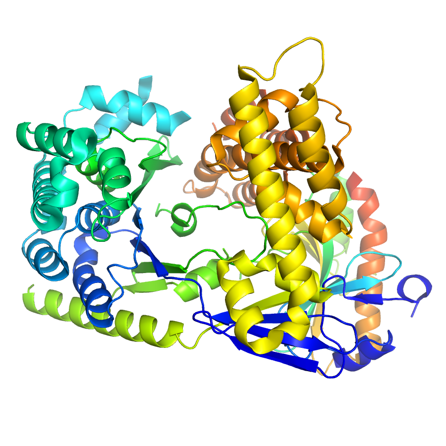
1.92 Angstrom resolution crystal structure of the full-length SpcU in complex with full-length ExoU from the type III secretion system of Pseudomonas aeruginosa
Edit deposit information
- CSGID target
- IDP91634
- PDB Id
- 3TU3 (NCBI MMDB)
- Authors
- 'A.S.Halavaty,D.Borek,Z.Otwinowski,G.Minasov,J.L.Veesenmeyer,G.Tyson,L.Shuvalova,A.R.Hauser,W.F.Anderson,Center For Structural Genomics Of Infectious Diseases (Csgid)'
- Responsible person
- Andrei Halavaty
- Responsible lab
- Northwestern University
- Deposition Date
- Sep 15, 2011
- Release Date
- May 23, 2012
Annotation
- Description
- Summary: Disease causing bacteria often manipulate host cells in a way that facilitates the infectious process. Many pathogenic gram-negative bacteria accomplish this by using type III secretion systems. In these complex secretion pathways, bacterial chaperones direct effector proteins to a needle-like secretion apparatus, which then delivers the effector protein into the host cell cytosol. Knowing the molecular details of this process is crucial to understanding pathogenesis. The effector protein ExoU and its chaperone SpcU are components of the Pseudomonas aeruginosa type III secretion system. Secretion of ExoU has been associated with more severe infections in both humans and animal models. The 1.92 Å resolution X-ray structure of the ExoU–SpcU complex was determined at the Center for Structural Genomics of Infectious Diseases (CSGID). This is the first structure of a full-length type III effector in complex with its full-length cognate chaperone. This crystallographic data provide a foundation for future studies aimed at designing inhibitors of this potent toxin. Methods: ExoU and SpcU were cloned, expressed, and purified from Escherichia coli. Catalytic activities of ExoU alone and in complex with SpcU were assessed in the presence of eukaryotic co-activators. The strength of the ExoU–SpcU interaction was evaluated by Surface Plasmon Resonance spectroscopy. The ExoU–SpcU complex was crystallized by the sitting-drop vapor diffusion technique. The X-ray structure of the complex was determined by the multiple isomorphous replacement method. Results: In solution, recombinant ExoU is catalytically active in the presence of eukaryotic co-activators. SpcU significantly reduces phospholipase activity of ExoU alone and in complex with the chaperone. The two proteins interact with a nanomolar affinity. ExoU has four distinct domains defined by differences in structure and function. SpcU interacts with three of the four domains of ExoU and defines the relative orientation of the ExoU domains. Solution and structural data shed light on oligomerization state of both proteins and the ExoU–SpcU complex. Conclusions: The toxin is catalytically inactive in the presence of SpcU and/or in the absence of host cell co-activator. The structure of the ExoU–SpcU complex is important in understanding how effector proteins are targeted to secretion into host cells. The structure allows interpretation of previous genetic and biophysical data. Although a structural model of an ExoU–co-activator complex is not available yet; it is proposed based on current and previous data that both SpcU and a co-activator may result in a similar relative orientation of the ExoU domains. In addition, an ExoU–host membrane model is discussed. This work provides an important tool for further understanding of the mechanism by which ExoU kills host cells.
- Functional assignment
- Toxin (Type III Effector Protein)/Toxin Chaperone
Ligands
| Ligand code | Name | Ligand type |
|---|
Structure information
Unit cell parameters
- Space Group
- C 1 2 1
- Unit Cell
-
a=154.14Å, b=52.58Å, c=119.54Å
α=90.00, β=126.59, γ=90.00 - Solvent content
- Matthews coefficient
- Resolution range
- 29.17-1.92Å (1.97-1.92Å)
- Rall(%)
- 19.3
- Rwork(%)
- 19.1 (22.5)
- Rfree(%)
- 22.5 (26.9)
- Num. observed reflections
- 58993 (4300)
- Num. Rfree reflections
- 2949 (212)
- Completeness(%)
- 99.8 (99.9)
- Num Atoms
- 5173
- Num Waters
- 490
- Num Hetatoms
- 501
- Model mean isotropic B factor
- 36.090Å2
- RMSD bond length
- 0.009Å
- RMSD bond angle
- 1.397°
- Filename uploaded
- 3TU3.pdb (uploaded on May 25, 2012 12:42 PM)
- Inserted
- May 25, 2012