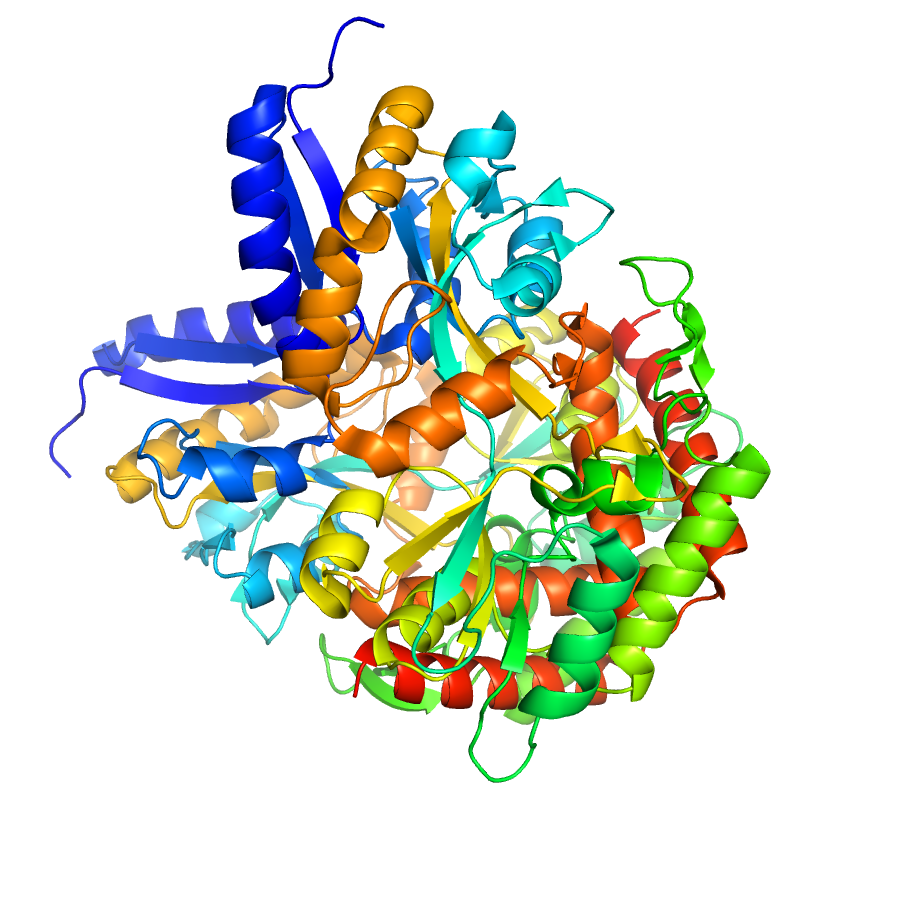
Structure of IDP91126
2.6 Angstrom Structure of the Extracellular Solute-binding Protein from Staphylococcus aureus in complex with PEG.
Edit deposit information
- CSGID target
- IDP91126
- PDB Id
- 4HS7 (NCBI MMDB)
- Authors
- 'G.Minasov,L.Shuvalova,I.Dubrovska,J.Winsor,F.Bagnoli,F.Falugi,M.Bottomley,G.Grandi,W.F.Anderson,Center For Structural Genomics Of Infectious Diseases (Csgid)'
- Responsible person
- George Minasov
- Responsible lab
- Northwestern University
- Deposition Date
- Oct 29, 2012
- Release Date
- Nov 07, 2012
Annotation
Ligands
| Ligand code | Name | Ligand type |
|---|---|---|
| P33 | 3,6,9,12,15,18-hexaoxaicosane-1,20-diol | crystallization |
| PEG | crystallization | |
| 175 | 3,5-dihydro-5-methylidene-4h-imidazol-4-on |
Structure information
Unit cell parameters
- Space Group
- P 65
- Unit Cell
-
a=101.86Å, b=101.86Å, c=191.34Å
α=90.00, β=90.00, γ=120.00 - Solvent content
- Matthews coefficient
- Resolution range
- 29.99-2.60Å (2.67-2.60Å)
- Rall(%)
- 17.0
- Rwork(%)
- 16.7 (22.9)
- Rfree(%)
- 21.9 (30.8)
- Num. observed reflections
- 34114 (2498)
- Num. Rfree reflections
- 1705 (121)
- Completeness(%)
- 99.0 (99.6)
- Num Atoms
- 6178
- Num Waters
- 291
- Num Hetatoms
- 356
- Model mean isotropic B factor
- 39.590Å2
- RMSD bond length
- 0.012Å
- RMSD bond angle
- 1.370°
- Filename uploaded
- rcsb075854.pdb (uploaded on Nov 12, 2012 3:38 PM)
- Inserted
- Nov 12, 2012