Structure of IDP91519
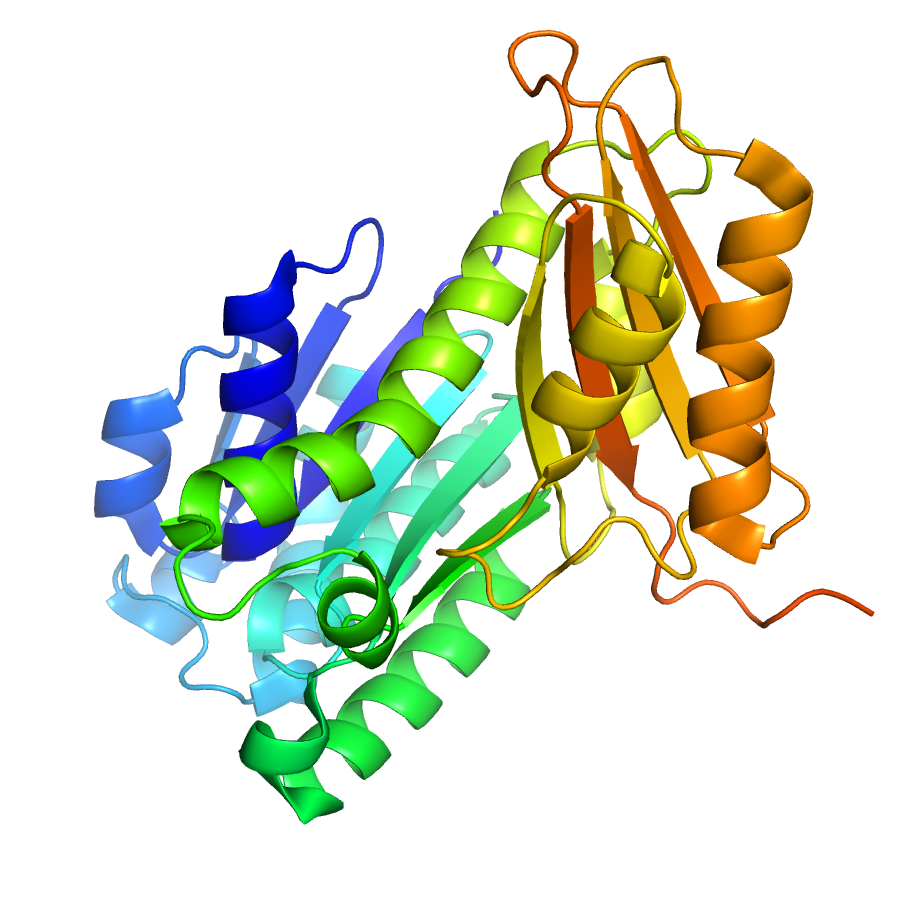
1.43 Angstrom resolution crystal structure of cell division protein FtsZ (ftsZ) from Staphylococcus epidermidis RP62A in complex with GDP
Edit deposit information
- CSGID target
- IDP91519
- PDB Id
- 4M8I (NCBI MMDB)
- Authors
- 'A.S.Halavaty,G.Minasov,J.Winsor,I.Dubrovska,E.V.Filippova,D.B.Olsen,A.Therien,L.Shuvalova,K.Young,W.F.Anderson,Center For Structural Genomics Of Infectious Diseases (Csgid)'
- Responsible person
- Andrei Halavaty
- Responsible lab
- Northwestern University
- Deposition Date
- Aug 13, 2013
- Release Date
- Sep 04, 2013
Annotation
- Description
- The crystal structure of the cell division protein FtsZ (ftsZ; construct with residues 2–392) from Staphylococcus epidermidis RP62A in complex with GDP was determined by molecular replacement and refined to 1.43 A resolution. The PISA and crystal packing analysis did not reveal any assemblies for the single chain of the protein in the C2 asymmetric unit. The tertiary structure of FtsZ consists of two separate domains. An N-terminal domain spans residues 2–222 and consists of a parallel 6-stranded beta-sheet that is flanked by 11 helices. A C-terminal domain is composed of an antiparallel 4-stranded beta-sheet and 2 helices that encompass residues 223–392. The protein is fairly acidic with theoretical pI of 4.8; the C-terminal domain has substantial surface patches of negative charge. Guanosine-5’-diphosphate is bound at a surface exposed pocket in the N-terminal domain. Residues 107–110 and 20–22 create an oxyanion hole that coordinates the diphosphate moiety. The nucleobase portion of the nucleotide is held in place via stacking interactions with Phe183 and hydrogen-bonded contacts with Asn25, Arg29 and Asn166. The sugar moiety interacts with Glu139, Arg143 and Asn166.
- Functional assignment
- Cell Cycle
Ligands
| Ligand code | Name | Ligand type |
|---|---|---|
| SO4 | sulfate | crystallization |
| 175 | 3,5-dihydro-5-methylidene-4h-imidazol-4-on | biological |
Structure information
Unit cell parameters
- Space Group
- C 1 2 1
- Unit Cell
-
a=71.33Å, b=52.06Å, c=87.00Å
α=90.00, β=109.64, γ=90.00 - Solvent content
- Matthews coefficient
- Resolution range
- 27.33-1.43Å (1.47-1.43Å)
- Rall(%)
- 17.1
- Rwork(%)
- 16.9 (32.8)
- Rfree(%)
- 20.6 (41.0)
- Num. observed reflections
- 53058 (3717)
- Num. Rfree reflections
- 2705 (187)
- Completeness(%)
- 95.8 (91.7)
- Num Atoms
- 2458
- Num Waters
- 333
- Num Hetatoms
- 374
- Model mean isotropic B factor
- 23.410Å2
- RMSD bond length
- 0.014Å
- RMSD bond angle
- 1.833°
- Filename uploaded
- 4M8I.pdb (uploaded on Jun 05, 2015 10:55 AM)
- Inserted
- Aug 29, 2013