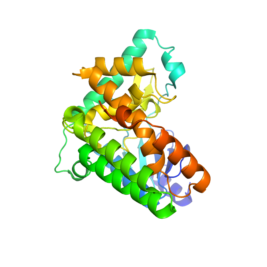
Structure of IDP91187
Crystal structure of aminoglycoside phosphotransferase APH(2")-Ib/APH(2'')-IIa in complex with ADP
Edit deposit information
- CSGID target
- IDP91187
- PDB Id
- 4DCA (NCBI MMDB)
- Authors
- P.J.Stogios,G.Minasov,A.U.Singer,K.Tan,B.Nocek,E.Evdokimova,O.Egorova,R.Di Leo,A.Savchenko,W.F.Anderson,Center For Structural Genomics Of Infectious Diseases (Csgid)
- Responsible person
- Peter Stogios
- Responsible lab
- University of Calgary
- Deposition Date
- Jan 17, 2012
- Release Date
- Feb 01, 2012
Annotation
- Description
- Aminoglycoside phosphotransferase (APH) enzymes act in a substrate- and position-specific manner and confer resistance to the activity of various aminoglycoside antibiotics. APH enzymes show a eukaryotic protein kinase-like fold with an insertion that takes part in substrate recognition. This is the ADP-bound structure of APH(2'')-Ib, an enzyme that acts on gentamicin, kanamycin, neomycin and other aminoglycosides.
- Functional assignment
- kinase
Ligands
| Ligand code | Name | Ligand type |
|---|---|---|
| ADP | biological | |
| MSE | modified residue | |
| 175 | 3,5-dihydro-5-methylidene-4h-imidazol-4-on |
Structure information
Unit cell parameters
- Space Group
- P 21 21 2
- Unit Cell
-
a=64.94Å, b=88.47Å, c=62.30Å
α=90.00, β=90.00, γ=90.00 - Solvent content
- Matthews coefficient
- Resolution range
- 29.38-1.80Å (1.86-1.80Å)
- Rall(%)
- 19.7
- Rwork(%)
- 19.5 (28.6)
- Rfree(%)
- 22.5 (33.3)
- Num. observed reflections
- 33681 (2742)
- Num. Rfree reflections
- 1687 (146)
- Completeness(%)
- 94.6 (99.0)
- Num Atoms
- 2449
- Num Waters
- 346
- Num Hetatoms
- 436
- Model mean isotropic B factor
- 45.810Å2
- RMSD bond length
- 0.009Å
- RMSD bond angle
- 1.147°
- RMSD dihedral angle
- 14.924°
- Filename uploaded
- 4DCA.pdb (uploaded on Feb 14, 2012 9:01 AM)
- Inserted
- Mar 23, 2011