Structure of CPX_06552_06553
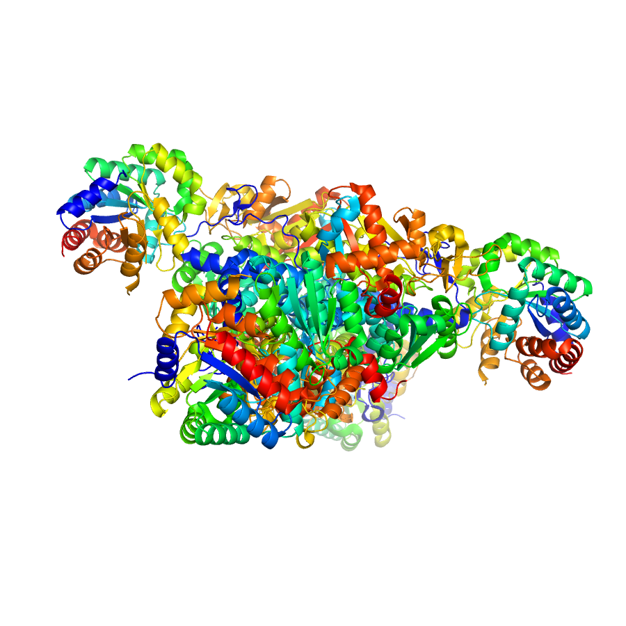
Crystal structure of tryptophan synthase from M. tuberculosis - AMINOACRYLATE- AND BRD6309-BOUND FORM
Edit deposit information
- CSGID target
- CPX_06552_06553
- PDB Id
- 6UB9 (NCBI MMDB)
- Authors
- Chang, C., Michalska, K., Maltseva, N.I., Jedrzejczak, R., McCarren, P., Nag, P.P., Joachimiak, A., Satchell, K., Center for Structural Genomics of Infectious Diseases (CSGID)
- Responsible person
- Changsoo Chang
- Responsible lab
- Argonne National Laboratory
- Deposition Date
- Sep 11, 2019
- Release Date
Annotation
Ligands
| Ligand code | Name | Ligand type |
|---|---|---|
| FMT | formate | crystallization |
| MLA | crystallization | |
| P1T | biological | |
| H9V | biological | |
| CS | biological | |
| ACT | crystallization | |
| PGE | crystallization | |
| EDO | ethylene diol | crystallization |
Structure information
Unit cell parameters
- Space Group
- P 21 21 21
- Unit Cell
-
a=135.37Å, b=159.62Å, c=164.93Å
α=90.00, β=90.00, γ=90.00 - Solvent content
- Matthews coefficient
- Resolution range
- 29.89-2.78Å (0.00-0.00Å)
- Rall(%)
- 17.0
- Rwork(%)
- 16.9 (0.0)
- Rfree(%)
- 21.0 (0.0)
- Num. observed reflections
- 85940 (0)
- Num. Rfree reflections
- 1710 (0)
- Completeness(%)
- 93.6 (0.0)
- Num Atoms
- 19206
- Num Waters
- 172
- Num Hetatoms
- 460
- Model mean isotropic B factor
- 43.120Å2
- RMSD bond length
- 0.000Å
- RMSD bond angle
- 0.000°
- Filename uploaded
- D_1000244280_model-annotate_P1.pdb (uploaded on Jun 23, 2020 4:12 PM)
- Inserted
- Jun 23, 2020